Chordae tendineae
From Wikipedia, the free encyclopedia
| Chordae tendineae | |
|---|---|
 |
|
| Interior of right side of heart. | |
 |
|
| The chordae tendineae are the unlabled, white tendons seen connecting the valves to the heart muscle | |
| Latin | chordae tendineae cordis |
| Gray's | subject #138 532 |
| MeSH | A07.541.510.240 |
| Dorlands/Elsevier | c_31/12236883 |
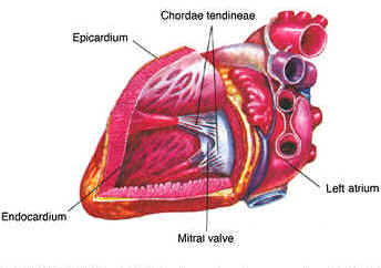
The chordae tendinae are cord-like tendons that connect the papillary muscles to the tricuspid valve and the mitral valve in the heart.
When the right ventricle of the heart contracts, the blood pressure pushes the tricuspid valve which closes and prevents a backflow of blood into the right atrium. The chordae tendineae prevents the flaps from being everted into the right atrium. Similarly, these cord-like tendons hold in position other flaps like the bicuspid or mitral valve.
[edit] External links
- SUNY Labs 20:st-0701
- Dictionary at eMedicine chordae+tendineae+of+heart
- Dictionary at eMedicine false+chordae+tendineae
- Organology at UC Davis Circulatory/heart/chambers0/chambers11 - "Mammal heart, chambers (Gross, Low)"
- Bioweb at UWLAX Human heart model (bicuspid valve)
- Illustration at health-pictures.com
| This cardiovascular system article is a stub. You can help Wikipedia by expanding it. |

